https://www.gosh.nhs.uk/news/new-study-pinpoints-why-some-transplanted-kidneys-are-rejected/
New study pinpoints why some transplanted kidneys are rejected
17 Sep 2025, 8:44 p.m.
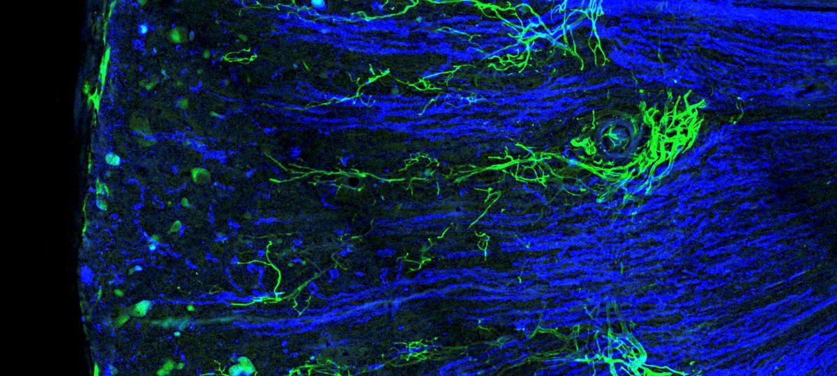
Kidney tissue with lymphatic vessels (green) and tubules (blue), captured using advanced 3D microscopy. The microscope that enabled this imaging was set up by Dr Dale Moulding at UCL Great Ormond Street Institute of Child Health, funded by the NIHR GOSH Biomedical Research Centre
Scientists have uncovered how lymphatic vessels – the kidney’s ‘plumbing system’ – undergo dramatic changes during chronic transplant rejection, becoming disorganised and spreading to unusual parts of the kidney. For the first time, this technique has allowed researchers to map lymphatics in the human kidney in 3D, revealing a delicate, tree-like network in the cortex, the region that filters blood and helps prevent the loss of vital molecules into urine.
Researchers at University College London (UCL), the Wellcome Sanger Institute and the University of Cambridge combined technology that determines the genetic or RNA sequence of individual cells with powerful 3D imaging to look at the lymphatic vessels in kidney tissue.
Published in the Journal of Clinical Investigation, the research sheds new light on one of the biggest challenges in kidney transplantation and could open the door to new treatments that help transplants last longer.
Kidney tranplants
The kidney is the most commonly transplanted organ in the UK. Figures from NHS Blood and Transplant show more than 8,000 people are on the waiting list so the more we can understand about rejection, the better.
Although the short-term outcomes of kidney transplantation – within a year after surgery – are very good, the long-term outcomes are poorer. Within 10 years, and depending on what country patients are treated in, roughly 50 per cent of kidney grafts will fail.
The researchers compared samples from both healthy and transplant rejection patients on a very large scale, to generate a huge amount of data. Then the team stained large chunks of kidney tissue whilst still intact and used a procedure to make it transparent. This 3D imaging helped prove the predictions from the genetic analysis of single cells.
The teams discovered that during transplant rejection, the lymph vessels change dramatically. They spread to deeper parts of the kidney known as the medulla, which does not normally contain lymphatic vessels.
Previously, researchers were unsure if the lymphatic system should be deemed as protective or detrimental in kidney transplants. This study suggests that it is normally protective but impaired in transplant rejection and so future research may focus on regenerating or recovering the lymphatic system in chronic kidney rejection.

Dr Daniyal Jafree, first author at the Wellcome Sanger Institute and an Honorary Research Fellow at UCL Great Ormond Street Institute of Child Health, said: “You can think of lymphatic vessels as the kidney’s plumbing system — clearing away excess fluid, immune cells and inflammation. Until now, we haven’t really understood what these vessels do in kidney transplantation because they are so difficult to study. Using new imaging techniques, we’ve shown for the first time that these vessels undergo dramatic changes during rejection.”

David Long, Professor of paediatric nephrology at UCL Great Ormond Street Institute of Child Health, and deputy theme lead of our NIHR GOSH Biomedical Research Centre, added: “Our innovative methods have allowed us to clearly demonstrate the important role of lymphatic vessels in transplant rejection.
“By combining single-cell sequencing with advanced 3D imaging, we’ve made a significant step forward in kidney transplant research.”
These findings challenge the view that lymphatic vessels do not participate in transplant rejection. Instead, the research suggests they change in ways that encourage rejection by altering their structure and fuelling immune responses. The results also pave the way for treatment research into targeting the lymphatic vessels to recover the system.
The amazing images you can see from this research were taken using equipment funded by our NIHR GOSH Biomedical Centre.
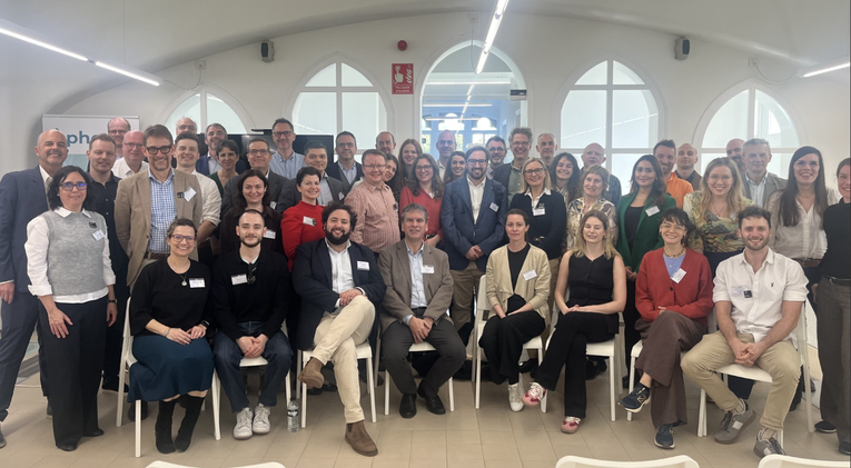
GOSH joins European health leaders to shape the future of paediatric data sharing
More than 50 clinicians, data scientists, digital health innovators and industry leaders came together in Barcelona last month to tackle one of the biggest challenges in paediatric healthcare: how to share health data safely across borders to improve care

Engineered tissue offers hope for children born with ‘missing’ food pipe
Scientists from Great Ormond Street Hospital (GOSH) and University College London (UCL) have created the first lab‑grown oesophagus - the food pipe - shown to safely replace a full section of the organ and restore normal function, including swallowing, in
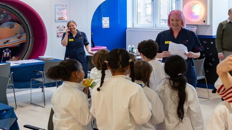
GOSH celebrates Rare Disease Day 2026 with local community
GOSH specialises in rare disease research and to celebrate, we recently invited two local North London Year 4 classes to join us for the day to learn about how we care for patients with rare diseases.
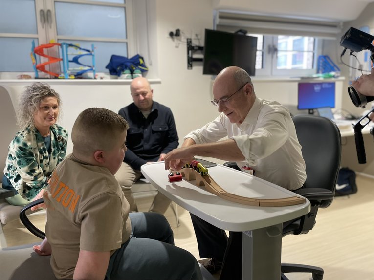
Professor Francesco Muntoni wins prestigious research prize
Professor Francesco Muntoni, GOSH Paediatric Neurology Consultant, has been awarded the 2026 Novo Nordisk Prize in recognition of his pioneering work transforming the outlook for children with Duchenne muscular dystrophy (DMD).