https://www.gosh.nhs.uk/news/mosaic-of-over-700-photos-wins-annual-moment-of-discovery-image-competition/
Mosaic of over 700 photos wins annual Moment of Discovery Image competition
27 Feb 2026, 7 a.m.
A beautiful collage made from over 700 photographs taken from across GOSH has been crowned the winner of our 5th annual Research and Innovation image competition ‘A Moment of Discovery’.
Staff across GOSH and our affiliated institutes, including the National Institute for Health and Care Research Great Ormond Street Hospital Biomedical Research Centre (NIHR GOSH BRC), University College London Great Ormond Street Institute of Child Health (UCL GOS ICH) and sites from our NIHR GOSH BRC Paediatric Excellence Initiative were invited to submit an image that captured ‘a moment of discovery’ in their work.
The winning image
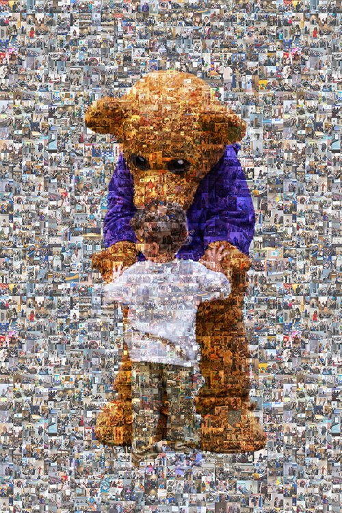
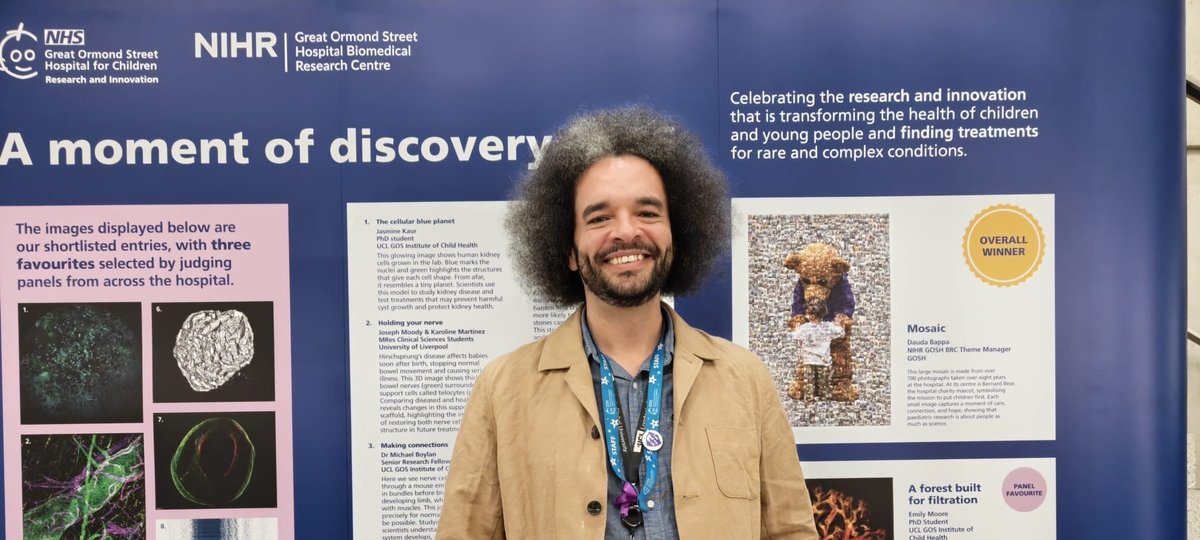
The overall winner: Mosaic - entered by Dauda Bappa, NIHR GOSH BRC Theme Manager Lead
The winning image ‘Mosaic’ was entered by Dauda Bappa, NIHR GOSH BRC Theme Manager Lead. The image is made from over 700 photographs taken over eight years at GOSH. At its centre is Bernard Bear, the GOSH Charity mascot, symbolising the mission to put children first. Each small image captures a moment of care, connection, and hope, showing that paediatric research is about people as much as science.
The entries ranged from microscopy to photography to scans, and offered snapshots of the research and innovation taking place across GOSH to find new treatments for rare or complex conditions, and transform the lives of seriously ill children and young people.
Dauda Bappa, winner of this year’s image competition said:
“Photography is my passion, and it’s a privilege to align that with my work at GOSH. My biggest challenge was condensing eight years and nearly 1,000 photos into a single impactful image.
“After talking to my wife and colleagues, the 'mosaic' concept clicked – to showcase the incredible breadth of people dedicated to improving children's lives and to champion the voice of the patient and public involvement community, we are building at GOSH.
“I’m genuinely honoured to be voted this year’s winner among such talent (shoutout to the Ghost entry!), and I’m very grateful to the NIHR GOSH BRC for the opportunity to capture these moments.”
Dr Kiki Syrad, Director of Research and Innovation at GOSH, said:
“Every year, the image competition gives us a window into the amazing research and innovation taking place across GOSH and our partners - it’s incredible to see such a variety of images. ‘Mosaic’ is a striking image and a wonderful reminder that groundbreaking research is built from many individuals.”
Professor Thomas Voit, Director of the NIHR GOSH BRC, said:
“Besides its artistic value, what makes this image so powerful is the way it brings together our values of inclusion and co‑creation. Every photograph that forms the ‘Mosaic’ represents someone contributing their time and expertise to supporting our shared purpose. These moments come together to reflect the spirit of collaboration at GOSH to help support our children and young people.”
Aoife Regan, GOSH Charity's Director of Impact and Charitable Programmes, said: "It's amazing to see the Charity front and centre in this beautiful image, highlighting the importance of collaboration when it comes to improving outcomes for seriously ill children. At GOSH, every moment matters as we look to create an environment that gives children the best chance, and best childhood possible. We'd like to say a big congratulations to Dauda, for being voted this year's winner."
Panel favourites
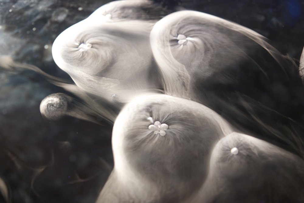
Ghost – entered by Zigfriedo Canada, Outpatient Receptionist. Dry ice is used in gene therapy packaging to safely transport medicines. This image captures dry ice reacting with water, producing thick, rolling fog. The effect happens because carbon dioxide gas is released and rapidly cools the surrounding air. A fan was used to create movement in the image, giving it an eerie, almost magical feel while demonstrating a simple but striking scientific reaction.
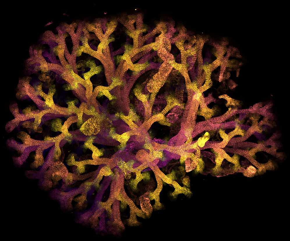
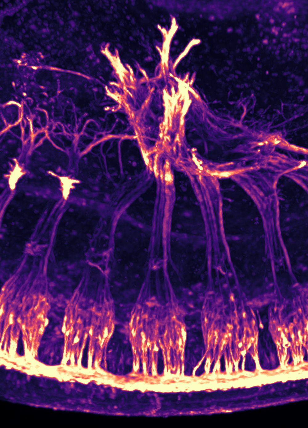
A forest built for filtration – entered by Emily Moore, PhD Student This image shows branching tubes in a developing mouse kidney. These collecting ducts form like a tree, growing outward from a central point to collect and carry waste to the bladder. The pattern mirrors kidney development in humans, helping scientists understand how healthy kidneys form and how this process can be disrupted by disease.
Shortlisted images
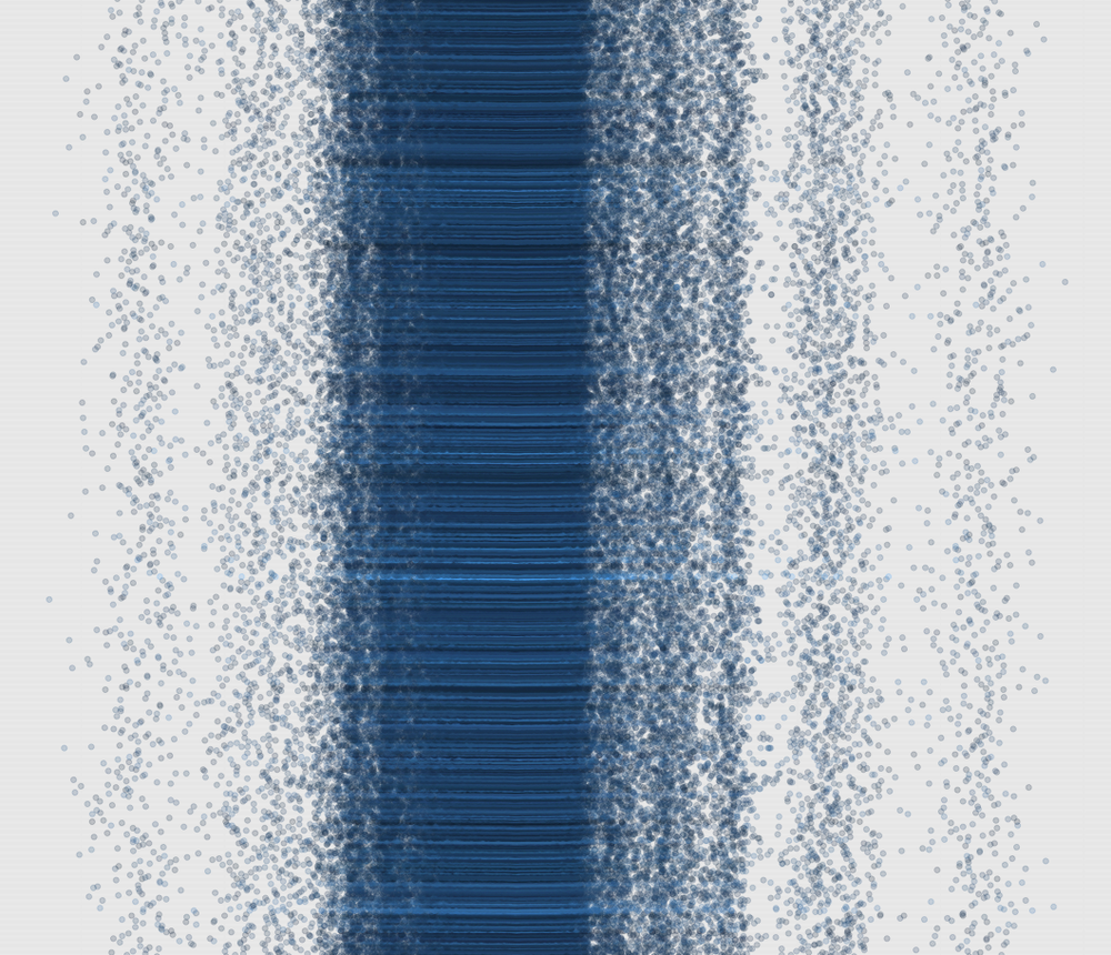
Ages and ages - entered by Dr Ramy Saad, Clinical Geneticist and Research Fellow This image comes from a study looking at how food related molecules in the body affect how our DNA ages. Each dot represents a person and a nutrient byproduct. When viewed all together, patterns emerge, showing how big data can feel overwhelming yet powerful. Like abstract art, meaning appears when you step back and change your perspective.
Making connections – entered by Dr Michael Boylan, Senior Research Fellow Here we see nerve cells migrating through a mouse embryo. They travel in bundles before branching into the developing limb, where they will connect with muscles. This journey must happen precisely for normal limb movement to be possible. Studying this process helps scientists understand how the nervous system develops, and what can go wrong in genetic conditions.
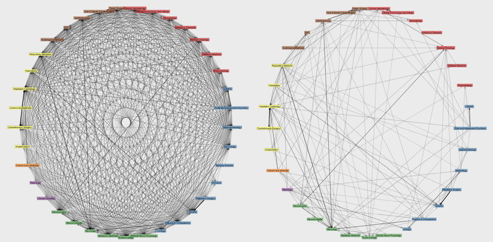
Complex Disease, Complex Hospital – entered by Dr William A Bryant, Data Science and Insights Lead This diagram shows how different medical specialties at the hospital are connected through shared patients with complex needs. Each line represents interactions during care, such as appointments or hospital stays. Zooming out reveals extensive collaboration across specialties, highlighting the importance of careful planning to deliver coordinated, efficient care for children with complex conditions.
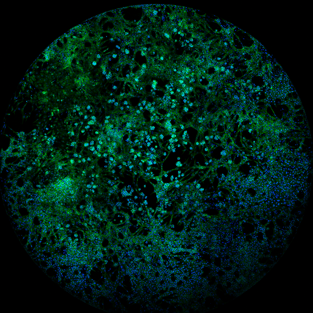
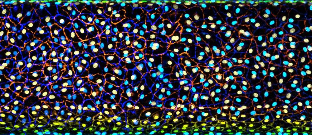
The cellular blue planet – entered by Jasmine Kaur, PhD Student This glowing image shows human kidney cells grown in the lab. Blue marks the nuclei and green highlights the cell structure that gives each cell shape. From afar, it resembles a tiny planet. Scientists use this model to study kidney disease and test treatments that may prevent harmful cell clumping and protect kidney health.
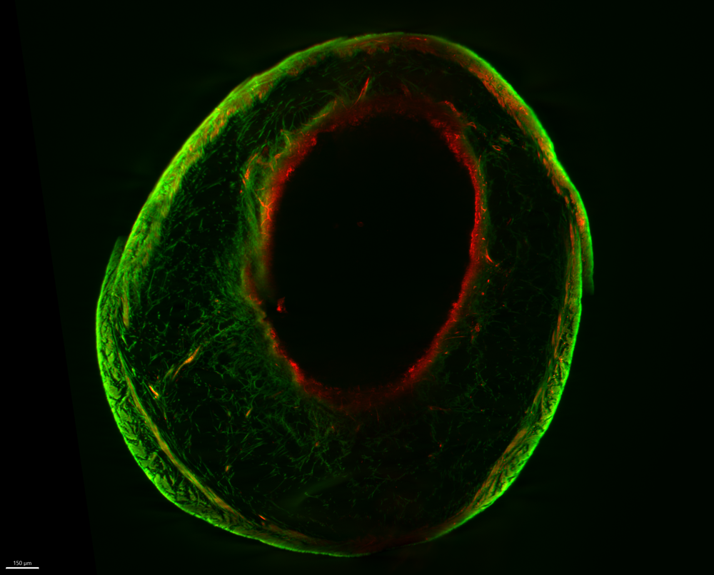
The Olive – entered by Dr Jess Trundle, Research Fellow This image shows a mouse bladder inflated to reveal its layered structure. Fluorescent dyes highlight different collagen networks that help the bladder stretch and contract. Studying this structure helps researchers understand bladder function and disorders, which is crucial for improving treatments for conditions affecting bladder control in children and adults.
Growing tiny tubes – entered by Dr Charlotte O’Riordan, Research Fellow This glowing image shows a lymphatic vessel grown in the lab. Hidden within childhood kidney cancers, these vessels can be used by cancer cells to spread. By recreating them outside the body, researchers can study how cancer hijacks these pathways and begin testing treatments to block this process and improve outcomes for children.
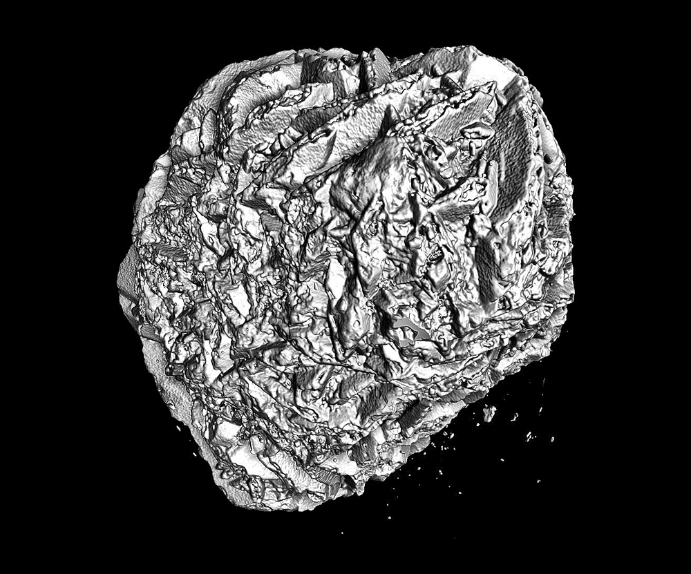
Attack of the stones – entered by Dr Aleksandra Berezowska, Research Fellow and Dr Ian Simcock, Clinical Academic Radiographer Kidney stones can form when tiny minerals in urine stick together and harden into crystals. Some children are more likely to develop them, and moving stones can cause pain and damage. This stone was removed at Great Ormond Street Hospital and scanned using MicroCT, revealing its structure within hours. This rapid insight may help prevent stones returning and support better treatments for children in the future.
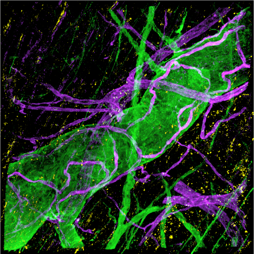
Holding your nerve – entered by Joseph Moody & Karoline Martinez MRes Clinical Sciences Students Hirschsprung’s disease affects babies soon after birth, stopping normal bowel movement and causing serious illness. This 3D image shows thickened bowel nerves (green) surrounded by support cells called telocytes (purple). Comparing diseased and healthy bowel reveals changes in this supporting scaffold, highlighting the importance of restoring both nerve cells and their structure in future treatments.
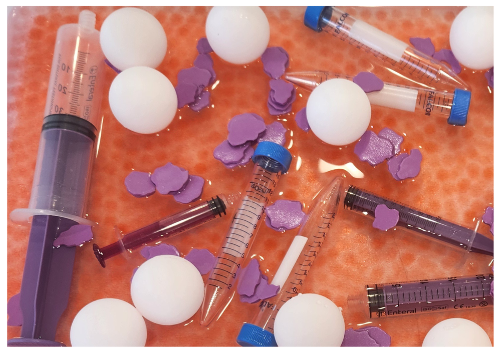
Hands on Haematology – entered by Kristen Black, Senior Play Specialist This photograph shows a sensory activity designed to help children learn about blood through play. Coloured beads and shapes represent red blood cells, white blood cells, plasma, and platelets. By exploring these materials handson, children can better understand blood tests and feel more confident, turning something scary into something familiar and fun.

Update for patients and families on industrial action
Some of our resident doctors at Great Ormond Street Hospital will be taking part in planned industrial action from 7am on Friday 14 November to 7am on Wednesday 19 November.

GOSH Voice to host Sibling Bowling Day for National Siblings Day
GOSH Voice will be hosting a sibling bowling day on 11 April 2026 to celebrate National Siblings Day on 10 April.
Engineered tissue offers hope for children born with ‘missing’ food pipe
Scientists from Great Ormond Street Hospital (GOSH) and University College London (UCL) have created the first lab‑grown oesophagus - the food pipe - shown to safely replace a full section of the organ and restore normal function, including swallowing, in
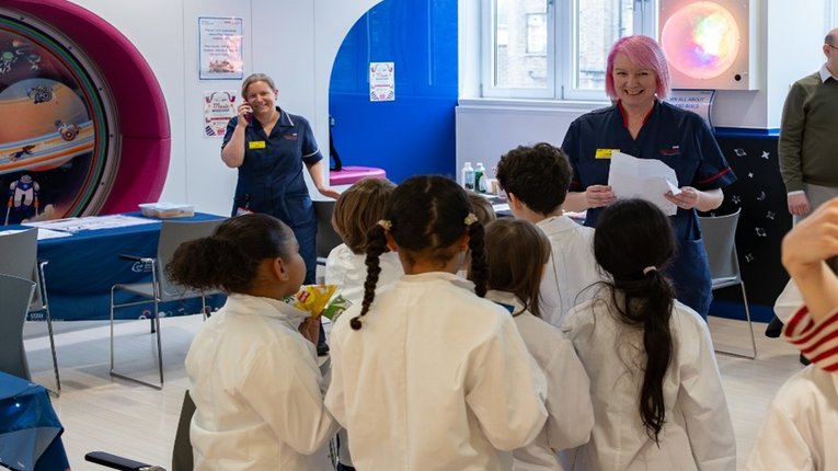
GOSH celebrates Rare Disease Day 2026 with local community
GOSH specialises in rare disease research and to celebrate, we recently invited two local North London Year 4 classes to join us for the day to learn about how we care for patients with rare diseases.