https://www.gosh.nhs.uk/news/a-moment-of-discovery-extraordinary-images-showcase-research-at-gosh/
A moment of discovery: extraordinary images showcase research at GOSH
29 Feb 2024, 9 a.m.
A vibrant image helping researchers' study gastrointestinal diseases and their potential treatments has been crowned the winner of the 3rd annual National Institute for Health and Care Research Great Ormond Street Hospital Biomedical Research Centre (NIHR GOSH BRC) image competition ‘A Moment of Discovery’.
Staff across GOSH and its affiliated institutes, the NIHR GOSH BRC and University College London Great Ormond Street Institute of Child Health (UCL GOS ICH) were invited to submit an image that captures ‘a moment of discovery’ in their research. The competition was also opened to other children’s hospitals across the UK within the NIHR GOSH BRC paediatric Excellent Initiative, including Alder hey, Birmingham and Sheffield.
11 images and gifs displaying the variety of research connected to GOSH were shortlisted and put before three panels – the GOSH Young Peoples Advisory Group for Research (YPAG), NIHR GOSH BRC stakeholders and the GOSH staff networks. These images offer glimpses into the research being undertaken at GOSH and its partners, that helps find new treatments for rare or complex conditions and hopes to transform the lives of seriously ill children and young people.
Voting across our three panels this year was incredibly close, and three favourite images were selected. These three favourites were put to the public who voted for an overall winner via social media.
After hundreds of votes across LinkedIn and Instagram, the image crowned the winner was ‘The explosive potential of gastrointestinal organoids’ entered by Giada Benedetti.
Overall winner: The explosive potential of gastrointestinal organoids

Image showing an exploding 'mini-organ' used to study gastrointestinal diseases and help find new therapies
Submitted by PhD student, Giada Benedetti, this image shows a gut ‘mini-organ’, known as an organoid, and it is a tiny copy of the digestive system. During a process used to visualise specific proteins, one of the organoids exploded, revealing its inner workings. These tiny organs are useful to model gastrointestinal diseases and are the perfect tool for scientists to study new therapies and test new drugs in the laboratory. In particular, the organoids can be derived directly from paediatric patients, and this gives us the opportunity to test therapies that specifically benefit the child.
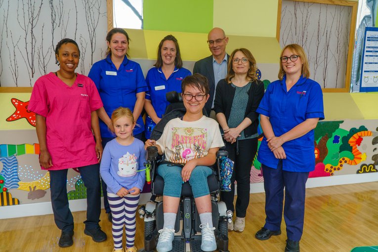
The team behind the image

Senior research associate Giovanni Giuseppe Giobbe, Marta Gazzaneo, Lucy Holland, and Giada Benedetti.
Pictured above are some of the team who helped create the winning image. The multidisciplinary team are based at the ZCR and are using these ‘mini-organs’ called organoids to study different diseases and use them to test new therapies.
“Sometimes it can be easy to lose sight of why we are performing research, but the close Interactions with the hospital really help motivate and give a clear purpose for the work that we do in the lab”
“The image competition is an excellent way to spread awareness for the research and display our scientific findings more dynamically and artistically, which is something that is easily lost in our usual world of numbers and spreadsheets”
Panel favourites:
It's about seeing the world

Image showing an embryonic eye stained for dystrophin
Submitted by PhD student, Reem Alkharji, this image shows the embryonic eye. The eye contains various specialised cell types which are crucial for sensing light and transmitting visual information to the brain. As development progresses, connections between the eye and the brain also develop, creating the optic nerve. Dystrophin proteins are required for normal visual functions. This image shows the embryonic eye stained with dystrophin to visualise its location and its contribution to the developmental journey. This work aims to discover what dystrophin and its associated proteins look like in developing brain and how it changes with development.
What we kneed to know.

Image showing the cells in a knee joint of a child with arthritis
Submitted by PhD student Chrissy Bolton, this image shows the cells in a knee joint of a four-year-old with arthritis. The red areas show the blood vessels infiltrating the tissue. They increase as the disease progresses, bringing in cells which attack the joint. The outer yellow and pink areas should be a thin barrier for the knee but becomes thicker as the joint tries to heal the damage caused by the inflammation. The green layers show scar tissue, which makes the joint stiffer and harder to move.
In the UK, inflammatory arthritis affects 1 in 1,000 children, typically affecting the large joints. After a decade of treatment, most children still suffer from ongoing disease. Most research in arthritis has focused on adults. How chronic inflammation differs in a child's developing immune system is still poorly understood. Ultimately, it was the parents who told us that their children must also benefit from the advances in technology that has driven research forward.
All shortlisted enteries
-

‘My lung is on fire’ was entered by PhD student Giuseppe Cala. This image shows a lung ‘mini-organ’, known as an organoid, grown from stem cells. It is a tiny, functional replica of a lung, mirroring its complexity. The blue shows the cell nuclei, the red shows the cell membrane, and the yellow/orange “fire” shows the moving hair-like structures called cilia. Lung organoids provide an up-close view of how the lungs work at a cellular level, they can be used to study various lung-related diseases and find potential therapies.
-

‘Lab is home’ was entered by Post-doctoral Researcher Maryam Clark. As a researcher you spend years working in the laboratory and moments of discovery are sometimes found alone at the microscope or late in the evening. Often, we only see the output of science and we rarely get to know the journey of the people behind the discoveries and how they felt whilst making them. “For many scientists, we are excited to share our discoveries with friends and families, which, for me, are my cats. With this illustration I wanted to combine my second home (the lab) and my family (my cats), showing my cats at my laboratory bench”.
-

‘The lights of life’ was entered by PhD student Atachapon Theppichaiyanond. During the development of the central nervous system (CNS), neural progenitors generate neurons by undergoing asymmetric division (cell division that generates a neuron and a neuron progenitor or two distinct neurons at the same time). In vertebrates this division is important to expand the neuronal population, produce neuronal diversity and shape the brain. Little research has been done on the molecular mechanisms that regulate asymmetric division and this research will enhance our understanding of vertebrate CNS development. This image shows a spinal cord of a zebrafish embryo with different neurons produced via asymmetric division. Confocal imaging identified the progenitor cells (green) that produce two distinct daughter cells (red and blue) via asymmetric division.
-

‘Syncytial formation of respiratory syncytial virus following cell infections’ was entered by PhD student Muhammad Pradhika Mapindra. Respiratory Syncytial Virus (RSV) is a respiratory virus that can cause mild flu like symptoms, but in infants and older adults it can be more severe and lead to hospitalisation with respiratory support. RVS is characterised by syncytia, which are formed by multiple cells fusing together. This image shows the syncytia after being exposed to RSV within five days. The green structures are RSV infected cells, and the blue circles show the cell nuclei.
-

‘Stabilised microtubules in zebrafish central nervous system’ was entered by PhD student Sara Anuar. Microtubules are tubular structures which form key components of the cytoskeleton in cells. They are essential for cell structure, intracellular transport, cell division and migration. Using spinning disc confocal microscopy, this image shows the rigid and unchanging tubular organisation of stabilised microtubules (blue) in the zebrafish central nervous system. Primary microcephaly is a rare disease where children are born with significantly smaller heads compared to population standards and usually result from defects on microtubules. By studying microtubules, we can understand the potential causes of primary microcephaly that contribute to the decrease of neural progenitor population and generate a smaller brain. Zebrafish can be used as a model to investigate the role of specific genes in microtubules and centrosomes of neural progenitor populations.
-

‘Insights into Down’s syndrome brainstem’ was entered by Post-Doctoral Researcher Ekin Ucunco. This image shows blood vessels (pink) and proliferative cells (blue/green) in the hindbrain of a foetus with Down’s syndrome. The hindbrain, formed by the cerebellum and brainstem, regulates many vital functions including heart rate and breathing. Proper development of the hindbrain requires tightly controlled processes. Imagery like this can help us to understand how these processes are regulated. We can also compare how this regulation differs in people with and without Down’s syndrome.
-

‘Glial cells in action’ was entered by Allied Health Professional Lucien Bonfante. This image shows glial cells (brown), nerve helper cells, which have long fibre like structures that adhere to blood vessels and transport nutrients and oxygen to surrounding nerve cells. The immunohistochemical staining of glial cells can be used for the diagnosis and classification of various conditions and tumours of the central nervous system.
-

‘3D modelling for safer neurosurgical planning’ was entered by 3D engineer Luke Smith. This giff shows a printhead moving left to right creating an accurate 3D printed model of a patient’s brain blood vessels. Arteriovenous malformations (AVM) are complex lesions that require extensive pre-surgical planning, and it is challenging for neurosurgeons to accurately visualise and deconstruct these structures. 3D printing is a promising tool for creating visual models of AVMs, helping surgeons to understand their structure and interactions with the surrounding brain. This helps enable safe surgical removal.
Lasting impressions
“It’s hard to believe that some of these images are of the human body!”.
“Quite a few phantasmic pictures, the exploding gastric organoid is sublime! The image competition is a fantastic visual representation of the power of investing in children’s research”.
“A beautiful set of images which show the breadth of work across GOSH and its affiliated institutes, these images often represent the first stages of improving the lives of children at GOSH and around the world. It is a reminder of how research and innovation is incorporated into everything we do at GOSH."
Showcasing our research
In celebration of Rare Disease Day on the 29th of February, all the shortlisted images will be exhibited at a research event in the ZCR. The image display will then tour the hospital, showcasing the research connected to GOSH to as many people as possible.
Landmark decision to screen for spinal muscular atrophy at birth
Great Ormond Street Hospital (GOSH) is delighted at the announcement that newborn babies in England will be offered screening for spinal muscular atrophy (SMA) through the newborn blood spot, or "heel prick", test programme.
From complex science to clear communication
Across multiple projects at GOSH, patients, families and young people are playing a vital role in transforming how research is communicated

Building the next generation of paediatric research leaders
From early career clinicians to emerging scientists, the NIHR GOSH Biomedical Research Centre (BRC)’s Academic Training Weekend is shaping the future of paediatric research - bringing together talent, expertise and ambition from across the UK.
A catalyst to unlock the next generation of research leaders
The NIHR GOSH Biomedical Research Centre (BRC) Fellowships are transforming early-career researchers into independent leaders - driving innovation, attracting millions in funding and shaping the future of paediatric science.